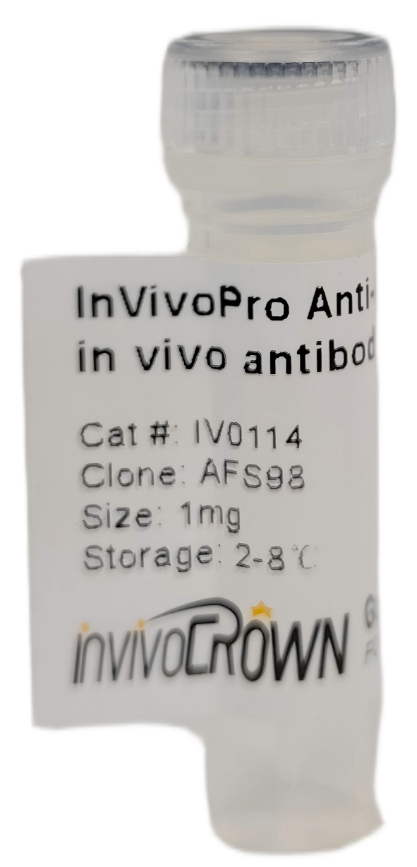
| Catalog |
IV0114 |
| Product Name |
InVivoPro Anti-Mouse CSF1R (CD115) in vivo antibody, Clone AFS98 |
| Size |
1mg/5mg/25mg/50mg/100mg |
| Isotype |
Rat IgG2a κ |
| Clone |
AFS98 |
| Target |
CSF1R |
| Other Names |
Macrophage Colony Stimulating Factor Receptor, M-CSFR, CSF-R1 |
| Isotype Control Catalog Code |
IV0105 or Rat IgG2a Isotype Control |
| Dilution Buffer |
PBS, pH 7.2, contains no stabilizers or preservatives |
| Reactivity |
Mouse |
| Host Species |
Rat |
| How much antibody to use in vivo |
400-500 μg per mouse; or 20 mg/kg. This range is based off the most recent publication data using the AFS98 clone in vivo. Each investigator should determine their own optimal working dilution for specific applications. |
| Background |
Macrophage colony-stimulating factor (M-CSF, also termed CSF-1) receptor is an integral membrane tyrosine kinase, expressed in monocytes (macrophages and their progenitors) and drives growth and development of this blood cell lineage. It also triggers growth and development of mononuclear phagocytes and placental trophoblasts. M-CSF receptor is present in osteoclasts both at mRNA and protein levels. When M-CSF ligand specifically binds to the M-CSF receptor on osteoclasts, it induces receptor dimerization, activation and autophosphorylation of cytoplasmic tyrosine residues M-CSF and its receptor present as candidate therapeutic targets in states of inflammatory bone erosion. |
| Applications |
In vivo macrophage depletion, in vivo monocyte depletion, Flow cytometry |
| Purification |
protein A or G |
| Storage |
This antibody is stable for at least 2 months when stored at 2-8°C. For long term storage, aliquot in working volumes without diluting and store at -20°C or -80°C. Avoid repeated freeze thaw cycles. |
| Shipping |
2-8°C with blue ice |
| Concentration |
Lot specific, generally ≥ 5.0 mg/ml |
| Shelf Life |
12 months from the date of receipt if stored as recommended |
| Formulation |
PBS Buffer, PH 7.2, with no carrier protein, or preservatives. |
| Sterility |
0.2 μM filtered |
| Endotoxin |
≤ 1.0 EU/mg, by the LAL method |
| Purity |
99% |